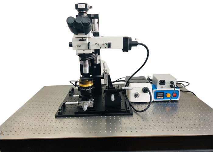
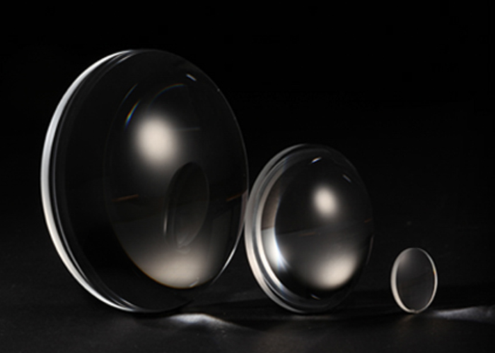
The more important function of a scanning electron microscope (sem) is to observe and analyze the microscopic morphology of samples. How to obtain a clear and practical image is the ultimate goal. In order to get a good image, the microscope objective supplier combined the existing data and summarized the following steps:
(1) At lower magnification, the sample to be analyzed, magnified, and focused samples can be found in the mobile sampling stage. The simpler method is to find out the characteristic points with obvious boundaries and use contrast, brightness, magnification, and focal length to make images. Be as clear as possible, and then look for or select interesting analysis sites based on samples.
(2) Rotate the sample stage or select the appropriate magnification by rotating the angle of the image group in the grating. In addition, in order to take good photos, we must not only explain the academic value of the problem but also try to consider the beauty of the panorama.

(3) On the basis of the original magnification, zoom in multiple times to make focusing and astigmatism more effective. At this time, you can choose to reduce the area to be focused. There will be a small area on the screen. In this small range, the scanning speed is faster, which helps to determine the incidence. Only by adjusting the focal length to the positive focal length, the low or over-focus of the incident beam can blur the image, eliminate rapid astigmatism every hour, and make the image details clear.
(4) Adjust the contrast and brightness of the analysis area of interest, so that the lining of the entire field of view can not only have clear black and white levels but also maintain proper contrast so that the surface level and details are rich. Otherwise, if the image is dim, reduce the details of the dark parts. Too much oversaturation or too dark will reduce the brightness, resulting in loss of detail.
When observing more than 10,000 times, it is necessary to eliminate astigmatism, and then focus on the focus. When the focus is low, the focus is too large, the vertical direction of the slender, the focus is not extended, but not obvious. Focus on the positive condition to adjust the time, position, and size of astigmatism until the image is clear. If the image is elongated, astigmatism is eliminated. At this time, the focus is completed due to scanning E. The focal length of the electron microscope changes magnification. Therefore, the focus time is generally 2-3 times the magnification, or a multiple of the number of observations, making it easier to determine whether it is positive. The focus knob used to adjust the image is a clearer focus. After moving the sample, the sample will reach a height.
If astigmatism or large-amplitude astigmatism device cannot change the amplitude or direction of astigmatism at a lower rate, you must consider whether the electron beam axis is damaged, whether the optical rod is seriously polluted, whether the sample is magnetic or obv, and whether there are impurities in the electron optical channel, or when there is a problem, it should be dealt with in a targeted manner for normal observation and analysis.
Our company provides 3D electric sliding table.


