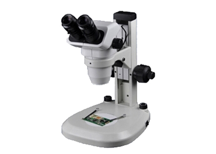
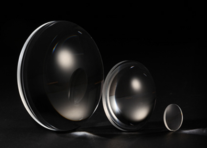
Pathological reading under the Microscope Objective is one of the important contents in the work of pathologists. Microscopic observation and recorded results are the scientific basis for clinical diagnosis. It is scientific to use the microscope with the correct standard to observe and record the results of the reading. The field of view (FN) of the microscope eyepiece is different. The size of the field of view can be seen under the microscope. Different area areas have an influence on the count of the positive rate under the microscope. We should understand the relationship between the eyepiece and the field of view. . The field of view that can be seen when the number of fields is small is small. Conversely, if the number of fields is large, the area of the field of view that can be seen is large.
1 Identification of the number of fields of view of the eyepiece
Microscope design and production have international standards, and the number of fields of view is identified on the eyepiece of the microscope. For example, the Olympus BX50 microscope, the number of fields of view of the eyepiece is 22 (the English and digital signs before the value of 22 are the eyepiece classification name and magnification).
2 Actual field of view and calculation formula
On the specimen plane, the area that the microscope can observe (circular area) is called the actual field of view, also called the field of view. The area size can be known by the following formula.
3 objective magnification
The objective lens is an important optical component for microscope imaging. Biomicroscopes commonly use objective magnifications of 4, 10, 20, 40, and 100. The high magnification objective used in pathology counting refers to 40 .
4 intermediate magnification
Direct observation through the eyepiece does not require consideration of the intermediate magnification. The intermediate magnification is the magnification of the CCD interface, photographic eyepiece, and CCD component added to the optical path. Because the microscopes used today are mostly infinity imaging systems, with the addition of fluorescence observation, differential observation, differential interference observation, etc., the components do not change the magnification, and need not be considered.
PDV is a China Microscope Objective Supplier. We have a high-tech work shop. We have a complete quality management system. Choose us, you can be get most careful and thoughtful service!


