Laser confocal microscope principle
Pulished on Dec. 11, 2019
Confocal laser scanning microscopy is a high-resolution microscopic imaging technique. When ordinary fluorescence optical microscopes observe thick specimens (such as cells), the fluorescence from the vicinity of the observation point will greatly interfere with the resolution of the structure. The key point of confocal microscopy technology is that only one point (focus) in space is imaged at a time, and then a two-dimensional or three-dimensional image of the specimen is formed by computer-controlled point-by-point scanning. In this process, light signals from out of focus will not interfere with the image, thereby greatly improving the sharpness and detail resolution of microscopic images.
The working principle of a general confocal microscope: a laser beam used to excite fluorescence is reflected by a dichroic mirror through a light source pinhole, and is converged by a microscope objective lens and incident At the focal point of the specimen put on the XY motorized microscope stage to be observed. The fluorescence (fluorescence light) generated by the laser irradiation is collected by the objective lens and sent to the dichroic mirror together with a small amount of reflected laser light. The fluorescence carrying image information has a relatively long wavelength, which directly passes through the dichroic mirror and passes through the detection pinhole to the photodetector (usually a photomultiplier tube (PMT) or an avalanche photodiode (APD) ), Into an electrical signal into the computer. Due to the dichroic mirror's spectroscopic effect, the residual laser light is reflected by the dichroic mirror and will not be detected.
The role played by the exit aperture: Only light emitted by points on the focal plane can pass through the exit aperture; light emitted by points outside the focal plane is defocused on the exit aperture plane, and most of them cannot pass through Small hole in the center. Therefore, the observation target point on the focal plane appears bright, while the non-observation point appears black as the background, the contrast is increased, and the image is clear. During the imaging process, the position of the exit aperture is always in a one-to-one relationship with the focal point of the microscope objective (conjugate conjugate, so it is called con-focal microscopy. Confocal display Microtechnology was invented by American scientist Marvin Minsky; he patented the technology in 1957. But it wasn't until the late 1980s that laser confocal scanning made significant progress in laser research due to significant advances in laser research Microtechnology (CLSM) has become a mature technology.

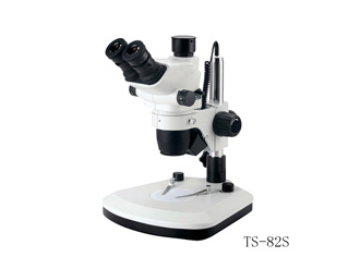
Stereo Microscope
Today's laser confocal microscopes have developed into a high-tech tool that combines a variety of cutting-edge scientific achievements such as laser technology, micro-optics, automatic control and image processing. It is one of the indispensable tools in modern micro research field. Adhering to the consistent corporate philosophy of "trust and creation", Nikon is providing the industry with world-leading confocal microscope system products.


